Human Skeletal System Diagram PDF | Printable Labeled Bone Anatomy Chart
Explore a detailed Human Skeletal System Diagram PDF with labeled bones, joints, and structure for biology, anatomy, medical education, and classroom learning.
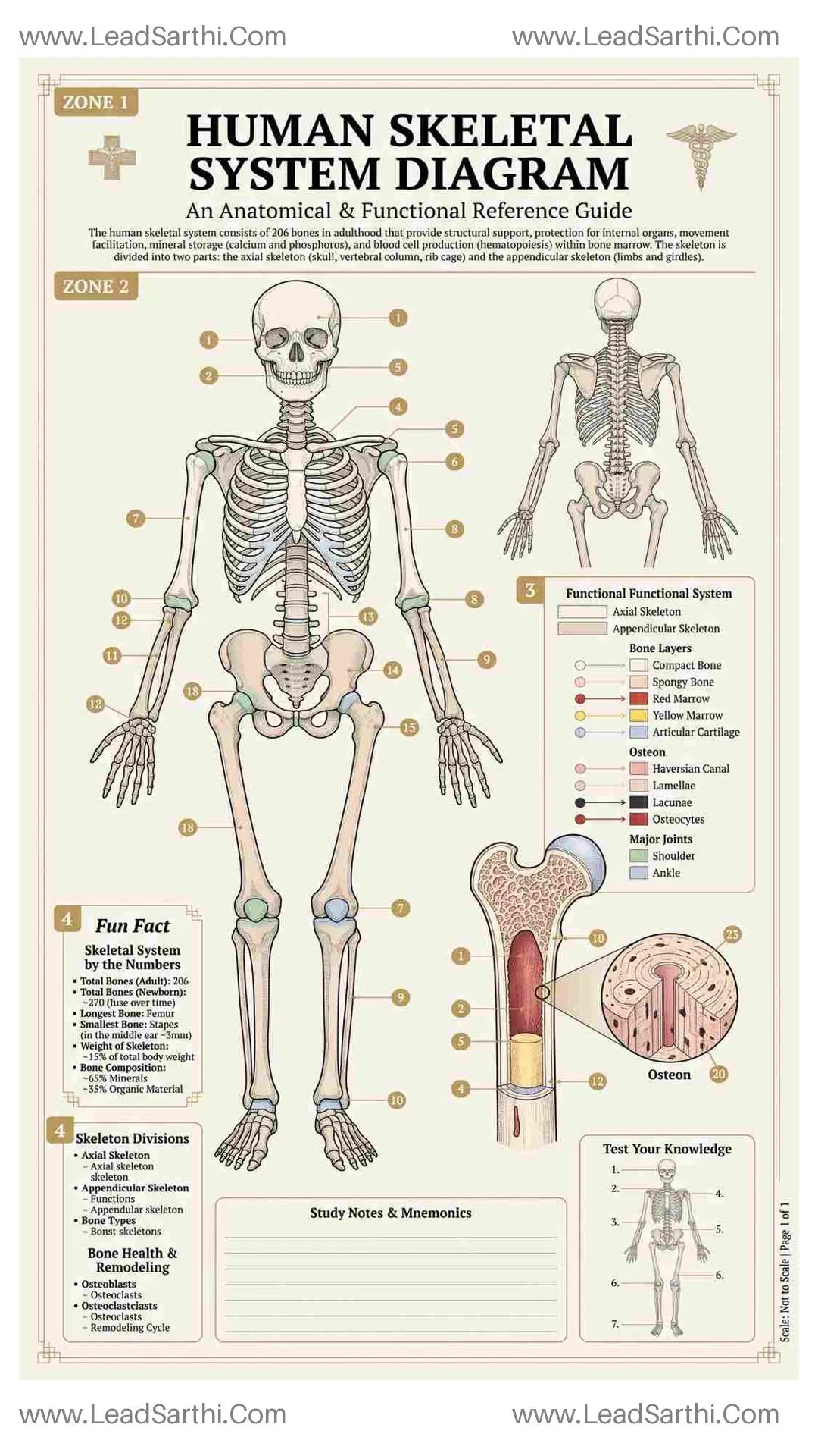
The Human Skeletal System Diagram is a fundamental educational resource for understanding the internal framework that supports and shapes the human body. The skeletal system provides structure, protects vital organs, enables movement, and stores essential minerals. A clear and well-labeled diagram helps learners easily visualize the arrangement and function of bones throughout the body.
The human skeleton is made up of 206 bones in adulthood, forming a strong yet flexible framework. Many students find it challenging to memorize bone names and understand how different parts of the skeleton connect. A detailed Human Skeletal System Diagram simplifies this by presenting a structured visual overview of the entire skeletal structure.
A typical diagram includes major bones such as the skull, clavicle (collarbone), scapula (shoulder blade), sternum, ribs, vertebral column (spine), pelvis, humerus, radius, ulna, femur, patella, tibia, and fibula. Some diagrams also highlight joints, cartilage, and key structural divisions such as axial skeleton (skull, spine, rib cage) and appendicular skeleton (limbs and girdles).
This file is available in a3, a4, a5, us letter and us half letter size format. Pick one from the following that you need.
By downloading any file, you agree to use it for personal purposes only. Do not redistribute, resell, modify, or share this file in any digital format.
More Similar Templates You May Like
Template #1#
Template #2#
Template #3#
Template #4#
Template #5#
Template #6#